
Figure 6.
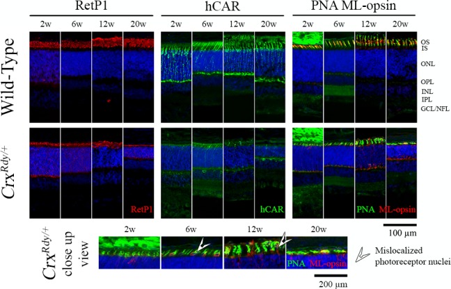
Immunohistochemistry using rod and cone markers. Frozen sections from the dorsotemporal retinal region of CrxRdy/+ kittens and WT controls at the indicated ages were immunostained with rhodopsin (RetP1), cone arrestin (hCAR), and medium/long wavelength-opsin (ML-opsin) along with the pan cone marker peanut agglutinin (PNA). The higher magnification views of CrxRdy/+ sections (bottom row) show mislocalization of ML-opsin to the inner segments, cell bodies, and pedicles of the cones (white arrowheads). ONL, outer nuclear layer; OPL, outer plexiform layer; INL, inner nuclear layer; IPL, inner plexiform layer; GCL/ NFL, ganglion cell layer/nerve fiber layer; white arrowhead, mislocalized photoreceptor nuclei.