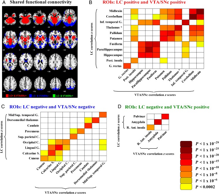
Figure 4.
(A) Brain areas that shared functional connectivity to the LC and VTA/SNc—positive connectivity (red color); negative connectivity (blue color); and negative connectivity to LC and positive connectivity to VTA/SNc (green color). (B–D) Matrices of R values of linear regressions (P < 0.05 corrected for multiple comparisons) between effect size (z scores) of ROIs with shared LC and VTA/SNc connectivity across all 250 subjects. There were 11 ROIs with PosR (red), 9 ROIs with NegR (blue), and 3 ROIs with negative connectivity to LC but positive connectivity to VTA/SNc (green). Asterisk: except dorsomedial thalamus; Inf.: inferior; G.: gyrus; Post.: posterior; R.: right; Ant.: anterior; Mid.: middle; Sup.: superior; C.: cortex; S.: sulcus.

