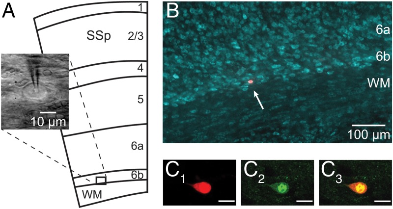
Figure 1.
Recordings in L6b. (A) Schematic representation (adapted from the Allen Mouse Brain Atlas; mouse.brain-map.org) of the different layers of the primary somatosensory cortex (SSp) and the underlying white matter (WM), together with an infrared microscopy image of a neuron recorded in this study. (B) Localization of an Alexa Fluor dye-injected neuron in layer 6b (arrow) with fluorescent Nissl counterstaining. (C1–3) Confocal snapshot images of a L6b cell recorded in a GAD67-GFP (line G42) transgenic mouse, showing Alexa Fluor dye-injected fluorescence (C1) and GFP fluorescence (C2) colocalized (C3) in the same cell. Scale bar in C1–3: 10 μm.

