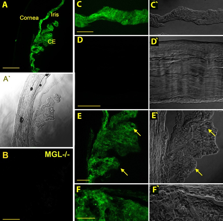
Figure 3.
Monoacylglycerol lipase is prominently expressed in ciliary epithelium and iris but not cornea. (A) Overview in a nonpigmented CD1-strain mouse shows MAGL expression in several major anterior eye structures. CE, ciliary epithelium. (A', C'–F') Corresponding differential interference contrast (DIC) images. (B) Monoacylglycerol lipase staining in tissue from MAGL KO anterior eye tissue, taken at same settings. (C) Iris is prominently labelled. (D) Monoacylglycerol lipase is not present in corneal epithelium, endothelium, or stroma. (E) Monoacylglycerol lipase is present in ciliary epithelium (arrow), particularly the inner pigmented layer. (F) Close-up of the angle shows prominent labelling of the ciliary body (lower portion of image) but little labelling of the trabecular meshwork. Scale bars: (A) 150 μm, (B) 50 μm, (C–E) 40 μm, (F) 25 μm.

