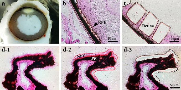
Figure 1.
Laser microdissection images of collected tissues from human and mouse eyes. (a) Fresh frozen section of human globe. (b) Human RPE tissue. (c) Human neural retinal tissue. (d1) Human ciliary process staining with Eosin Y, NPE (pink); (d2) NPE is selected in red; (d3) following laser microdissection of NPE.

