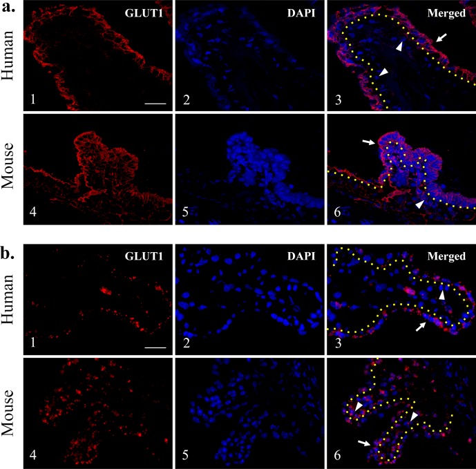
Figure 5.
Glucose transporter 1 immunofluorescence staining in human and mouse ciliary processes. Glucose transporter 1 expression was found predominantly at the NPE layer of human ciliary processes (a1–3), whereas mouse GLUT1 expression was present in both NPE and PE layers (a4–6). (b) Glucose transporter 1 in situ hybridization in human and mouse ciliary processes. The GLUT1 expression appears to be limited in the NPE as compared with the PE in humans (b1–3). Both NPE and PE layers reveal GLUT1 expression in the mouse ciliary processes (b4–6). Yellow dotted lines: separate PE and NPE; arrows: NPE; arrowheads: PE. Scale bar: 25 μm.

