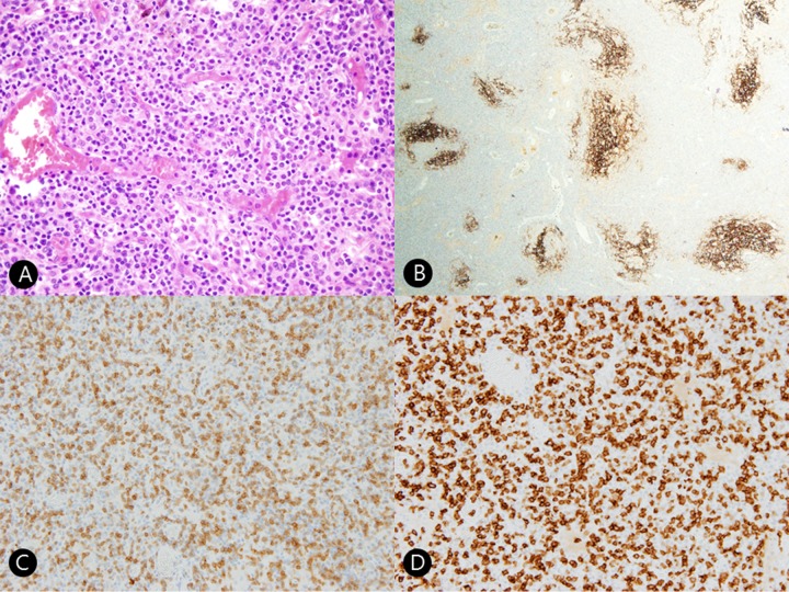
Figure 2.
(A) Histopathologic findings revealed intermediate-sized neoplastic T-cells with clear cytoplasm and proliferation of high endothelial venules (hematoxylin and eosin ×100). (B) Immunohistochemical staining for CD21 revealed arborizing follicular dendritic cells in a low-power view (CD21 immunostain ×40). (C) Tumor cells are positive for CD3 (×200). (D) Tumor cells are positive for CD4 (×200).

