Figure 3.

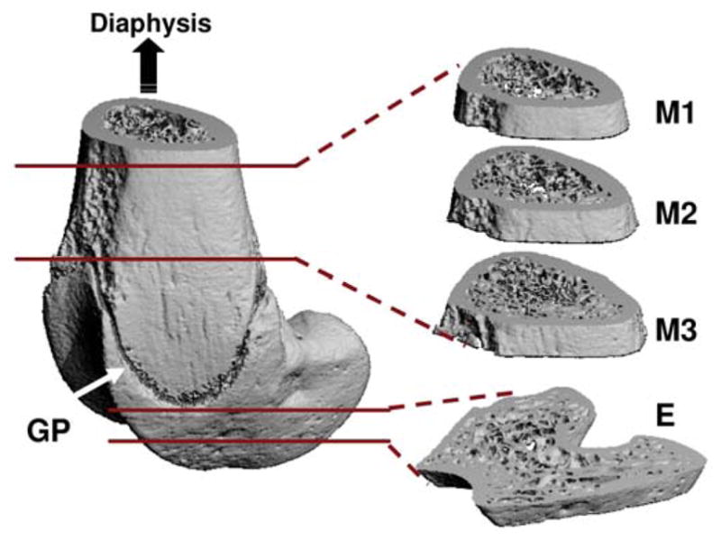
Trabecular bone at three metaphyseal sections and one epiphyseal region of the distal femurs was evaluated using microcomputed tomography. GP=growth plate (arrow); M=metaphysis; E=epiphysis. (Courtesy of BONE 2008;43:1093–1100).

Trabecular bone at three metaphyseal sections and one epiphyseal region of the distal femurs was evaluated using microcomputed tomography. GP=growth plate (arrow); M=metaphysis; E=epiphysis. (Courtesy of BONE 2008;43:1093–1100).