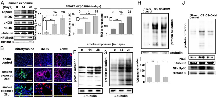
Fig. 2.
Tobacco smoke activates Rtp801 and consequent NF-κB–induced overexpression of iNOS and oxido-nitrosative stress in the lung. Immunoblots showing the levels of Rtp801, iNOS, eNOS, nNOS, and NF-κB–p65 in the whole-lung lysates of guinea pigs administered 14 and 28 d of CS exposure against air-exposed sham controls (A). (B) Representative profiles showing the relevant immunostained lung sections treated with specific antibodies followed by fluorescently tagged secondary antibodies against nitrotyrosine (green/Alexa Fluor 488), iNOS (red/Alexa Fluor 568), and eNOS (red/Alexa Fluor 568). (Scale bars: 100 µm.) (10 fields, n = 6.) Total NOS activity (C) determined in the above whole-lung lysates along with total nitrite (nitrate+nitrite) concentration in BALF (D) (measure of total NO released in the lung). Level of ROI (E) generated by freshly collected whole-lung lysates was also spectrofluorimetrically estimated by CM-H2DCFDA assay and shown in terms of relative fluorescence unit (rfu). Immunoblot shows the level of protein nitration (F) and oxidation of lung proteins (G) with increasing CS exposure (n = 6). Immunoblot showing the levels of protein oxidation (H), protein nitration, iNOS, and NF-κB–p65 expression (J) along with cellular ROI levels (I) in the lungs of animals exposed to 14 d of CS with or without DXM administration (nebulization), against air-exposed sham controls. Data were statistically analyzed by paired Student’s t test. Significant differences (**P < 0.01, ***P < 0.001) were observed in comparison with unexposed controls. Data are represented as means ± SD and represent three independent experiments done under similar conditions.

