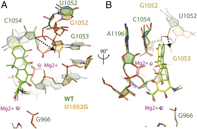
Fig. 3.
Structural perturbations to tetracycline binding to U1052G mutant ribosomes. (A) Superposition of the tetracycline-bound crystal structures of the wild-type and U1052G mutant ribosome. The dotted arrow represents the displacement of the phosphate group of nucleotide G1053 in the U1052G mutant. (B) This panel is rotated 90° along the vertical axis. wild-type is green, and the U1052G mutant is yellow. Magnesium ions are shown as pink spheres.

