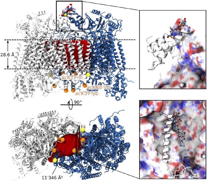
Fig. 5.
The lipid plug of CcO. Side view (Upper) and bottom view (Lower) of the CcO dimer (PDB ID code 2OCC). The membrane spanning hydrophobic region (28.6 Å) is indicated (Upper). The volume of the inner cavity (11′346 Å3, red) allows incorporation of seven or eight phospholipids or three or four CL, respectively. PTMs (phosphorylation, yellow and acetylation, orange) in the dimeric interface of CcO were located in the crystal structure. The bottom view of the complex reveals at least six acetylated lysine residues at the dimer interface, suggesting interactions with lipids. Protein–protein interactions in the dimer interface are shown as backbone and cartoon representation for the two monomers (magnifications). Amino acids in direct contact with other amino acids are shown as licorice, revealing a weak dimer interface stabilized by phospholipids.

