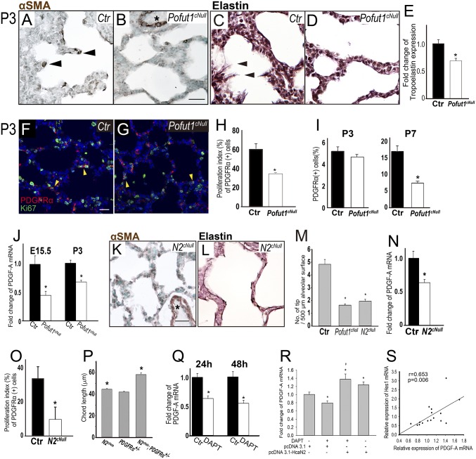
Fig. 3.
Inactivation of epithelial Notch signaling inhibits proliferation of mesenchymal AMYF progenitors. (A and B) Decreased number of αSMA-positive AMYFs (arrowheads) in (B) Pofut1cNull lungs compared with control (A) at P3. Asterisks indicate vascular SM. (C and D) Elastin staining revealed continuous elastin fibers with protrusions into the saccular space (arrowhead) in control (C). In contrast, only few protruding elastin fibers can be seen in Pofut1cNull lungs (D). (E) Tropoelastin mRNA significantly decreased in Pofut1cNull lungs. (M) Tip numbers of secondary septa significantly decreased in both Pofut1cNull and Notch2cNull lungs. (F and G) Proliferating AMYF progenitors are detected by PDGFR-α and Ki67 double staining of controls (F) and Pofut1cNull (G) lungs. The proliferation index estimated from the double staining was significantly reduced in Pofut1cNull and Notch2cNull lungs (H and O). (I) The number of PDGFR-α–positive AMFY progenitors was significantly decreased in Pofut1cNull lungs at P7, but not P3, during alveologenesis. (J and N) Real-time PCR showed significantly decreased PDGF-A mRNA expressions in Pofut1cNull (J) and Notch2cNull (N) lungs. AMYF differentiation (K) and elastin deposition (L) were also defective in Notch2cNull lungs. (P) Quantitative analysis revealed significantly increased chord length in Notch2cHete;PDGFR-α+/− mice at 3–6 mo old. DAPT (10 μM) inhibited PDGF-A mRNA expression in MLE15 cells (Q), and constitutively activated Notch2 signaling by transfection with HcaN2 plasmids rescued this phenomenon (R). Strongly positive correlation between levels of Hes1 and PDGF-A mRNA in MEL15 cells (S) (n = 3–5 in each group for quantification, *P < 0.05). (Scale bars, 10 μm.)

