Fig. S2.
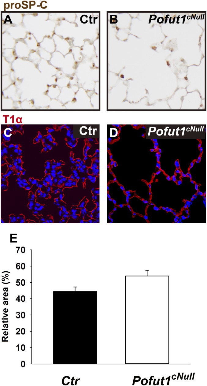
Distal lung differentiation in Pofut1cNull mice. Immunohistochemistry of (A and B) SPC, which labeled type II cells and of (C and D) T1-α (T1a), which labeled type I cells, showed a similar pattern of staining in control and Pofut1cNull lungs. (E) Quantification of type I cells presented as T1a staining area.

