Fig. 4.
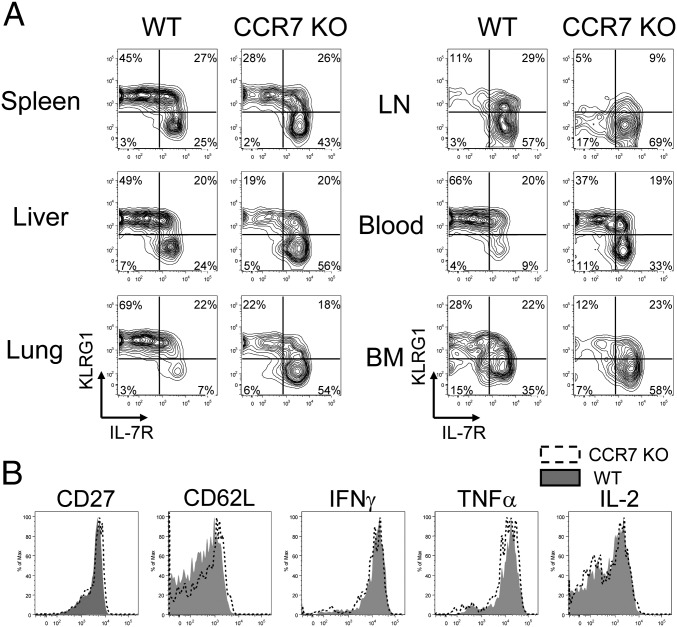
Phenotype differs between CCR7 WT and KO memory T cells. (A) On day 35 pi, P14 CCR7 WT and KO memory T cells were analyzed for the expression of IL-7R and KLRG1 by flow cytometry. (B) Memory T cells from the spleen were also analyzed for expression of CD27, CD62L (l-selectin), TNFα, IFNγ, and IL-2. Histogram plots show the expression of CD27 and CD62L on CCR7 WT (shaded) and KO (open), directly ex vivo. The production of IFNγ, TNFα, and IL-2 by CCR7 WT (shaded) and KO (open) cells, was assessed by 5-h GP33–41 peptide stimulation in vitro. All plots are gated on donor Ly5.1+ P14 CD8 T cells. Similar results were obtained from four independent experiments containing at least two mice per group.

