Fig. 5.
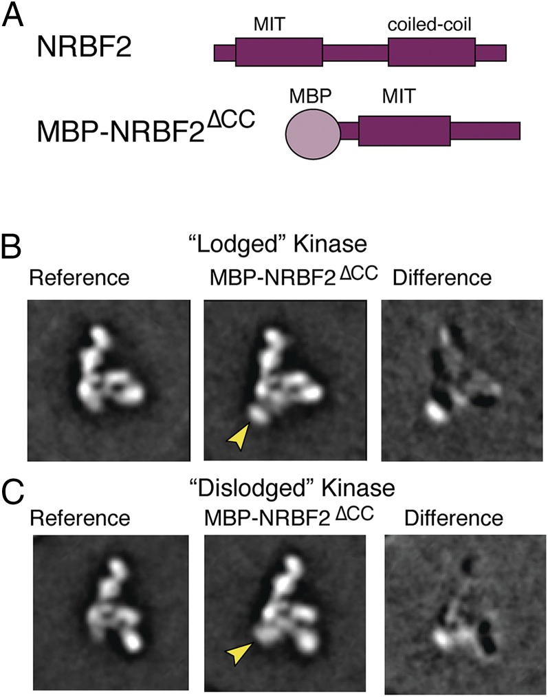
Negative-stain EM mapping of the NRBF2 binding site. (A) Cartoon of full-length NRBF2 and cartoon of MBP-tagged NRBF2∆CC. (B) (Left) EM 2D reference projection group of PI3KC3-C1 with kinase domain of VPS34 in the lodged position. (Middle) Two-dimensional projection groups of PI3KC3-C1 with MBP-NRBF2∆CC bound. Arrows indicate additional density. (Right) Two-dimensional difference maps showing density for MBP-NRBF2∆CC. (C) (Left) EM 2D reference projection group of PI3KC3-C1 with kinase domain of VPS34 in the dislodged position. (Middle) Two-dimensional projection groups of PI3KC3-C1 with MBP-NRBF2∆CC bound. Arrows indicate additional density. (Right) Two-dimensional difference maps showing density for MBP-NRBF2∆CC.

