
FIGURE 5.
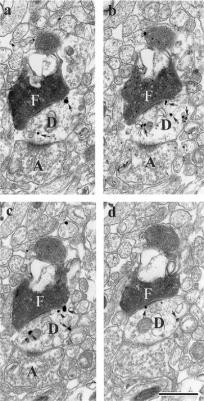
Series of electron micrographic sections through the nucleus reuniens of the midline thalamus (a–d) showing asymmetric contacts of a single PHA-L-labeled (F) fibers from the medial prefrontal cortex onto a labeled dendritic shaft, identified by the presence of numerous silver intensified gold deposits (arrows in D) of a RE cell retrogradely from a Fluorogold injection in the ventral hippocampus. Note also the presence of asymmetric contacts of an unlabeled fiber (A) on the same labeled dendrite segment (D). Scale bar = 1 μm.