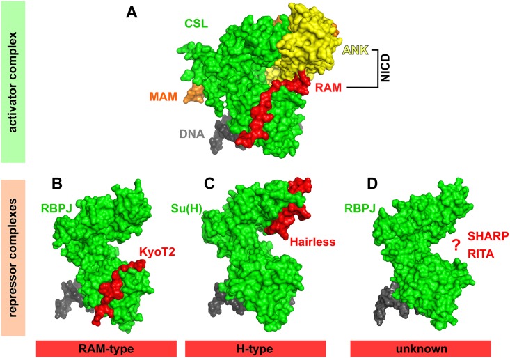
Fig 2. Surface views of the CSL coactivator complex (upper) and corepressor complexes (lower).
(A) The DNA-bound CSL activator complex consists of CSL (green), NICD (RAM domain, red; ankyrin repeats, yellow), and mastermind (MAM, orange). (PDB-ID: 1TTU). (B) KyoT2 (red) interacts with the BTD of CSL, similar to the NICD RAM domain (RAM-type). (PDB-ID: 4J2X). (C) Hairless interacts with the CTD of Su(H), resulting in a dramatic change of CTD conformation (H-type). (PDB-ID: 5E24). (D) The crystal structure of the SMRT/HDAC1 associated repressor protein (SHARP)-CSL corepressor complex and the CSL-RBPJ interacting and tubulin associated (RITA) corepressor complex is unknown at the moment (PDB-ID, RBPJ bound to DNA: 3BRG).

