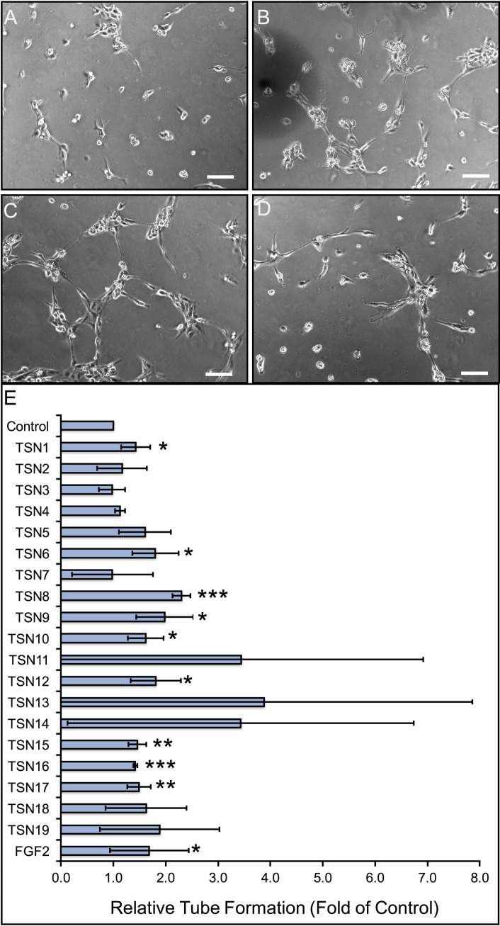
Fig 4. Quantitative analysis of peptide-induced dermal microvascular remodeling in vitro.
(A-D) Representative photomicrographs of human dermal capillary endothelial cell morphogenesis following treatment with 1% BCS (A), 100 nM TSN1 (B), 100 nM TSN 8 (C), and 100 nM TSN 10 (D). Scale bar represents 100 μm in all panels. (E) The lengths of human dermal capillary endothelial sprouts cultured on growth factor-reduced Matrigel in the presence or absence of 100 nM ECM-derived peptides were measured as described in “Methods” at 5 hours post-plating, with physiologic doses of FGF2 (0.6 nM) serving as positive controls. Data are displayed as fold increase in tube length, relative to 1% BCS-treated negative controls, and error bars represent standard deviation. * p < 0.05, ** p < 0.01, *** p < 0.001, compared with negative control (unpaired Student’s T-test).

