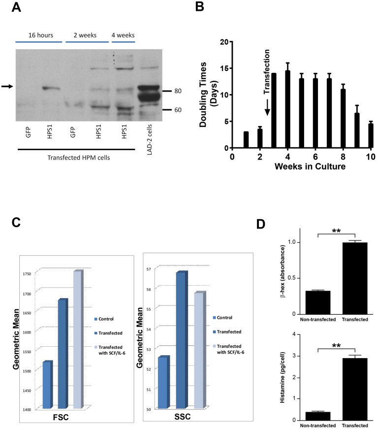
Fig 4. Results of transfection of HPM cells with normal HPS1.
A) Representative Western blot of HPS1 transfected HPM cells and control LAD2 cells demonstrating HPS-1 protein expression (arrow) in 16 hr, 2 and 4 wk HPM cultures, confirming transfection and expression of HPS-1 protein. Immunoblot is representative of at least 2 independent experiments; B) Slowing of doubling time 5–7 fold immediately following transfection with HPS1. Transfected cells reached average doubling rate of 3–4 days by 10 wks; C) FSC and SSC changes in HPM cells 4 wks following transfection. No additional changes were noted following supplementation of cultures for 4 wks with rhSCF and rhIL-6. Data are representative of 2 separate experiments performed in duplicate; D) Three-fold increase in cellular β-Hex content following transfection (upper graph) and ten-fold increase in cellular histamine content 4 wks following transfection (lower graph). Data are the means ± SEM (n = 3). **p<0.01.

