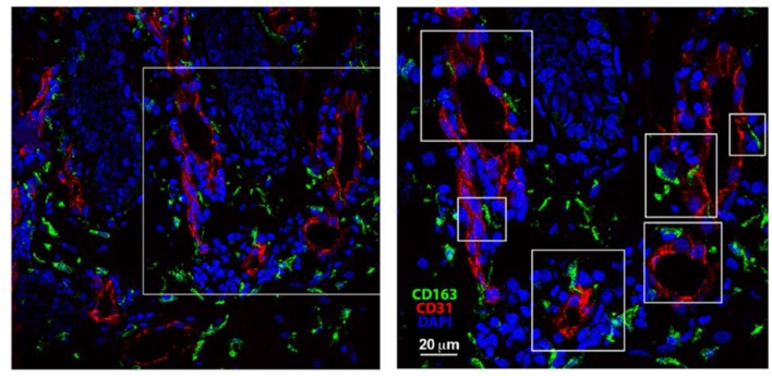
Author response image 1. Skin biopsies were collected, immediately embedded in OCT and frozen.
Then, the tissue was cut in 30 μm-thick criosections. After fixation and permeabilization, skin sections were blocked with human-γ globulin and 5% FBS. Then, sections were incubated with mouse anti-human CD31 for 1 hr followed by incubation with Alexa-Fluor 555 donkey anti-mouse. Next, samples were blocked with mouse serum 1:50 O/N 4°C, subsequently mouse-anti human biotinylated anti-CD163 at 5 μg/ml was added for 1 hr and, finally, samples were incubated with Streptavidin Alexa-Fluor 488. DAPI staining was performed just before mounting the sample for microscopy observation using a standard confocal microscope.

