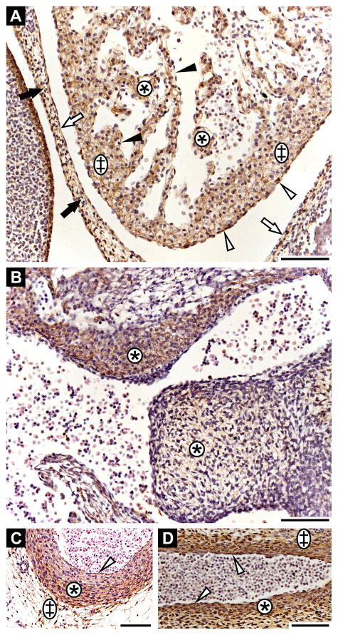
Figure 3. Fibrillin-3 expression in the cardiovascular system.
A: Heart anlage (6th GW). Fibrous pericardium (black arrows); parietal layer of serous pericardium (white arrows); visceral layer of serous pericardium (white arrowheads); spongy layer (asterisks); mantle layer of myocardium (double crosses); endothelium (black arrowheads). Note that the strong staining on the left originates from periderm. B: Endocardial cushion (6th GW; asterisks). C: Descending aorta at the level of the tracheal bifurcation in cross-section (8th GW). Tunica intima (white arrowhead); tunica media (asterisk); tunica adventitia (double cross). D: Descending aorta in longitudinal section (6th GW). Tunica intima (white arrowheads); tunica media (asterisk); tunica adventitia (double cross). Scale bars = 100 μm.

