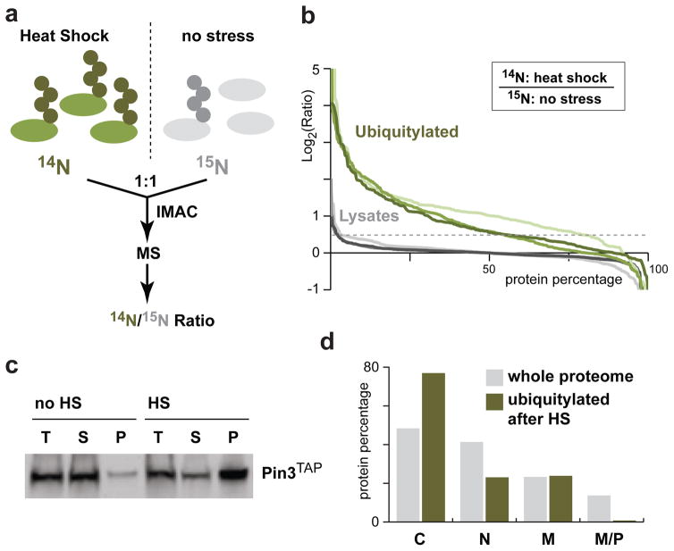
Figure 2.
Heat-shock mainly affects cytosolic proteins. (a) A schematic diagram of the workflow of the quantitative mass spectrometry analysis. (b) Percentage of proteins above the corresponding log2 values of the 14N heat-shock/15N no stress ratios for three independent experiments (I: light; II: medium; and III: dark). Analysis of proteins in the total cell lysate (grey: I, 486; II, 730; and III, 399) and of IMAC enriched ubiquitylated proteins (green: I, 302; II, 481; and III, 219) are shown. Proteins with a log2 ratio ≥ 0.5 are considered heat-shock affected. (c) Pin3TAP solubility was assessed prior to and after a 15 min 45°C heat-shock. An equal portion of each fraction (T: total; S: supernatant; P: pellet) was loaded on a SDS-PAGE for Western blot analysis with the anti-TAP antibody. (d) Subcellular localization of 155 proteins affected by heat-shock (green; identified as enriched in at least two of the three experiments in b) compared to the whole proteome (grey) - cytosol (C), nucleus (N), membrane (M) and mitochondria or peroxisome (M/P). Note that several proteins localize to more than one compartment.

