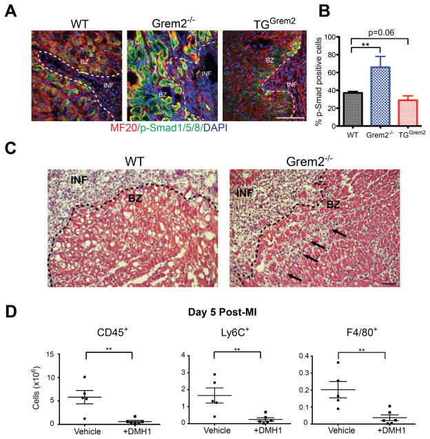
Figure 6. Grem2 regulates canonical/p-Smad1/5/8 BMP signaling in peri-infarct area cardiomyocytes.
(A) IF analysis of cardiac tissue sections 7 days post-MI using antibodies recognizing p-Smad1/5/8 (green) and Myosin Heavy Chain (MF20, red) shows activation of canonical BMP signaling in cardiomyocytes in the peri-infarct border zone. DAPI marks cellular nuclei. The number of p-Smad1/5/8+ cardiomyoctyes is increased in Grem2−/− mice and decreased in TGGrem2 mice. Scale bars 100 μm. BZ=infarct border zone; INF=infarct. (B) Quantification of p-Smad1/5/8+ cells in the infarct border zone as percentage of total MF20+ cells per viewing area between WT, Grem2−/−, and TGGrem2 mice. ** P < 0.01. Student’s two-tailed unpaired t-test. N=3. All data are means ± SEM. (C) Hematoxylin & Eosin staining 5 days post-MI shows that inflammatory cell infiltration (arrows) beyond the infarct border (dotted line) is greater in the Grem2−/− mice compared to WT counterparts. Scale bars 10 μm. BZ=border zone, INF=infarct. (D) Grem2−/− mice were injected once daily via IP with either the canonical BMP signaling inhibitor DMH1 or vehicle (DMSO) 2 days, 3 days, and 4 days post-MI. Number of total CD45+, Ly6C+ and F4/80+ cells in whole hearts at 5 days post-MI were determined by flow cytometry. DMH1 injected mice have a decreased inflammatory cell infiltration compared to vehicle controls. ** P < 0.01. Student’s two-tailed unpaired t-test. Vehicle N=5, +DMH1 N=6. Bars represent means ± SEM.

