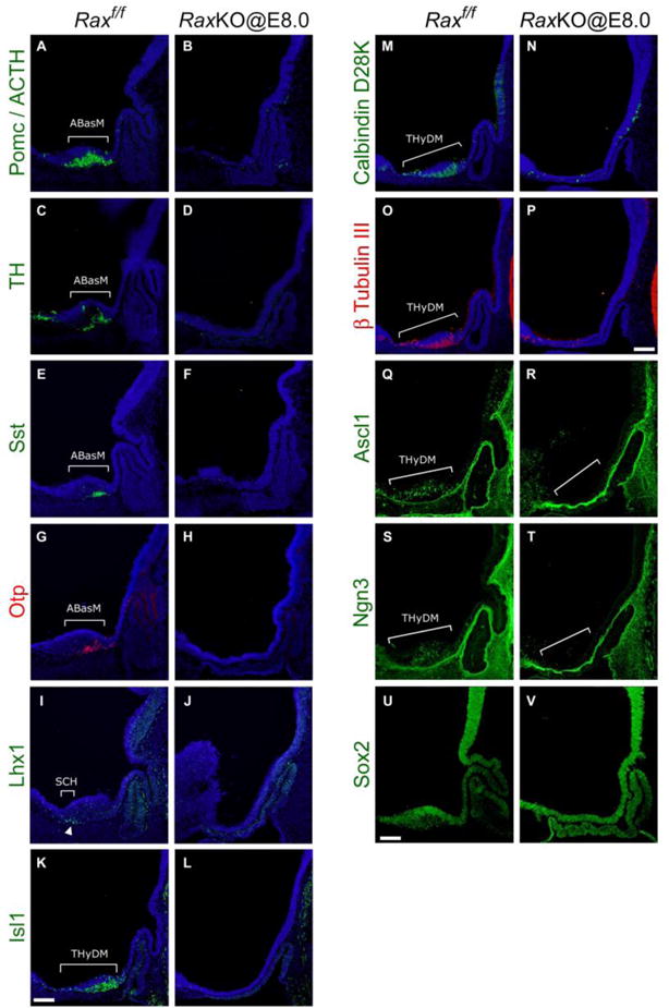
Figure 4.

Early loss of Rax eliminates dorsomedial hypothalamic marker expression. Immunohistochemical analysis on midsagittal sections of control (left on each panel) and RaxKO@E8.0 (right on each panel) mouse embryos. Proteins detected were: (A–B) Pomc/ACTH, (C–D) TH, (E–F) Sst, (G–H) Otp, (I–J) Lhx1, (K–L) Isl1, (M–N) Calbindin, (O–P) β-tubulin III, (Q–R) Ascl1, (S–T) Ngn3 and (U–V) Sox2. Embryos were collected either at E12.5 (Pomc/ACTH, TH, Sst, Otp, Lhx1, Sox2) or E11.5 (Isl1, Calbindin, β-tubulin III, Ascl1, Ngn3). Brackets indicate gene expression domains in the SCH, ABasM and THyDM. Note that expression of all marker genes in these domains is greatly reduced or eliminated in mutant embryos. Scale bars 100 μm.
