
Figure 5.
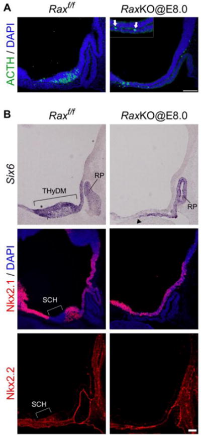
Effect of the loss of Rax function on hypothalamic transcription factors and neuron distribution. (A) Pomc/ACTH immunohistochemistry and DAPI staining of control (left) and RaxKO@E8.0 (right) embryos at E11.5. Several Pomc/ACTH neurons leave the thin neuroepithelium and relocate to the neighbouring oral ectoderm and mesenchyme, as shown in the inset of the right panel (arrows). (B) In situ hybridisation (Six6) and immunohistochemistry (Nkx2.1, Nkx2.2) analyses on midsagittal sections of control (left) and RaxKO@E8.0 (right) mouse embryos analysed at E12.5 (Six6, Nkx2.1) or E11.5 (Nkx2.2). In the control embryo, Six6 is expressed in the whole THyDM while Nkx2.1 is broadly expressed in the hypothalamus except for the alar area of the THyDM that corresponds to the future SCH, where Nkx2.2 is expressed. Elimination of Rax causes the loss of Six6 expression from the THyDM and loss of the characteristic gene expression (Nkx2.1−; Nkx2.2+) in the SCH domain. Note that Six6 expression in the Rathke’s pouch (RP) region extends dorsally in the oral ectoderm of the mutant (arrowhead; compare to Fig. 2). Scale bar 100 μm.