Figure 2.

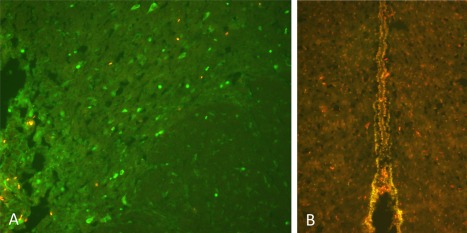
a. BrdU labeling of proliferative cells immediately surrounding the site of HFS‐DBS in the STN. b. Colabeling of Sox2 (green) and BrdU (red) positive cells around the third ventricle in STN‐HFS animals.

a. BrdU labeling of proliferative cells immediately surrounding the site of HFS‐DBS in the STN. b. Colabeling of Sox2 (green) and BrdU (red) positive cells around the third ventricle in STN‐HFS animals.