
Figure 4.
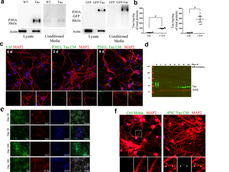
Tau from mouse primary neurons and human iPSCs can transfer via the extracellular medium (a) Lysate from rTg4510 primary neurons and P301L-GFP transduced neurons, and conditioned media from the same cells, labeled with TauC (full-length blots shown in Supplementary Figure 4). Actin shows equal amounts of protein loaded. (Full-length blots shown in Supplementary Figure 4.) (b) ELISA using tau-specific antibodies showing uptake of tau from media by tau-KO cells. Compared to controls (n = 4 cultures), tau was significantly higher in neurons after 1–6 hours of incubation with tau-conditioned media (n = 6, z = −2.558, adjusted P = 0.021) (left), and also after 1–6 days of incubation (n = 4, z = −2.309, adjusted P = 0.042). (c) Neurons (wild-type), incubated with rTg4510 conditioned media for 2 and 6 days (d), labeled with anti-tau antibody (green) and anti-MAP2 (red). Insets show tau in cell bodies and neurites. (d) Total tau in human iPSC neuron lysates. Recombinant tau ladder (R) separates in the following order of decreasing molecular weight: 2N4R, 2N3R, 1N4R, 1N3R, 0N4R, 0N3R. The single tau band corresponds to the 0N3R isoform. (e) iPSC cultures immunolabeled with the early forebrain marker Pax6 and tau, which was only expressed in post-mitotic neurons. By day 100 of differentiation, the majority of the cells in culture were immunoreactive for total and 3R tau. Scale bar, 100. (f) Neurons (wild-type) treated for 6 d with tau conditioned media collected from iPSC neurons, stained with antibodies (anti-tau, green, anti-MAP2, red) and DAPI (blue). Enlarged insets show tau (green) accumulating inside neurites (yellow). Scale bar, 10 μm.