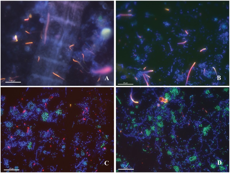
FIGURE 7.

Fluorescence in situ hybridization images of the WP microbial mat community at 630X magnification hybridized using the DSM-651 Desulfuromusa-specific probe. (A) Large DAPI-stained filament (blue) and chains of Desulfuromusa rods (red). (B) DAPI-stained cells (blue) and Desulfuromusa (red) emerging from a central axis and chains. (C) DAPI-stained filaments and cocci (blue) with gammaproteobacterial cocci (green) and Desulfuromusa (red). (D) Gammaproteobacterial cocci clusters (green) with DAPI-stained filaments (blue) and Desulfuromusa cells (red).
