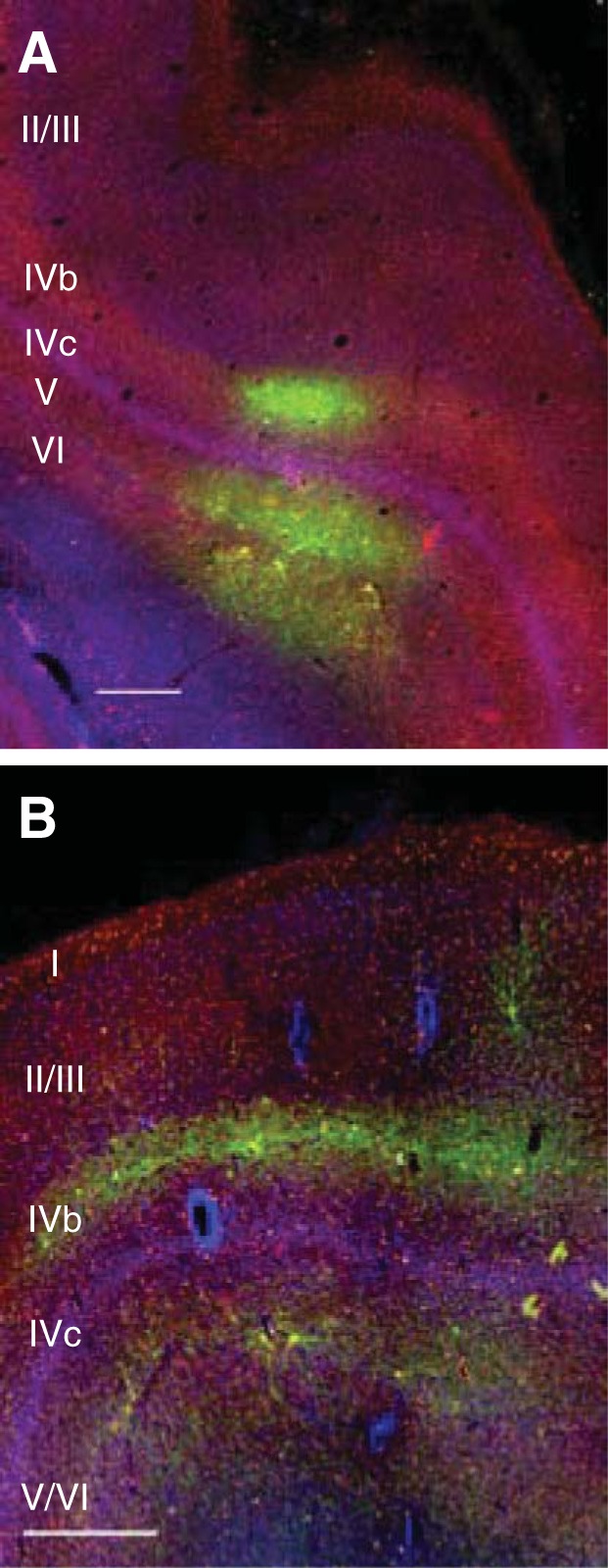
Fig. 1.

Viral vector tropism in primate neocortex. Coronal sections of visual area V1 of a rhesus macaque. Tissue was stained for SMI-32 (red) and with DAPI (blue), and transduced neurons are green. A: injection of LV-CaMKIIa-ArchT-GFP titered at 1.8 × 10⁷ infectious units/ml. B: injection of AAV9-hSyn-ChR2-eYFP titered at 2.75 × 10¹³ genomic copies/ml. A total of 5 μl of each viral vector was injected (1 μl injected at each of 5 sites, spaced 500 μm apart). Scale bars, 500 μm. The sparse labeling is consistent with some studies (Gerits et al. 2015) but not others (Diester et al. 2011).
