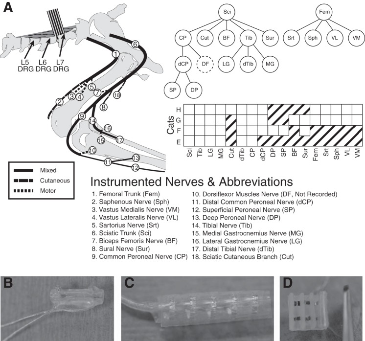
Fig. 1.
A: schematic of nerve cuff location in the left hindlimb. B: most nerves were instrumented with bipolar split-cuff electrodes. C: the femoral and sciatic trunks were each implanted with 5-contact cuffs, while all other nerves received bipolar cuffs. D: in experiment H a custom book electrode was implanted on three femoral nerve branches. The common peroneal and tibial nerves were both implanted with a proximal cuff, close to the initial branch point, and a distal cuff. The ankle dorsiflexor nerves tended to be deep and could not be implanted without significant dissection of the limb, but activity could be inferred by differential activation of the proximal and distal portions of the common peroneal nerve.

