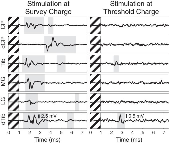
Fig. 4.

Compound action potentials recorded on distal branches of the sciatic nerve at the survey amplitude (left) and at the lowest amplitude at which any response was detected (right). Detected responses are highlighted in gray. This example highlights a DRG electrode (same as Fig. 3) that, at high amplitude, recruited many of the nerves innervating the ankle and foot but at low amplitude was selective for only the nerves projecting to the plantar surface of the paw. Hashed regions denote 1-ms blanking period.
