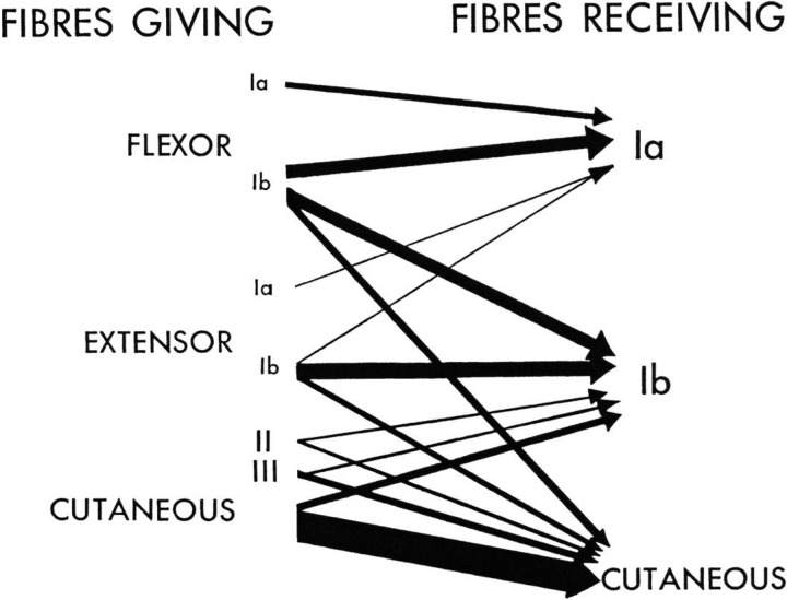
Fig. 3.
Classic diagram of Eccles et al (1963) showing the relative amounts of depolarization evoked in different types of primary afferent fiber by other afferent fibers; the thicker the arrow, the greater the depolarization and hence the degree of presynaptic inhibition. Note that, in the case of jaw muscles (Goldberg and Nakamura 1977), as well as in limb muscles (Rudomin and Schmidt, 1999), there is now evidence that cutaneous fibers can produce PAD in group I afferents. Reproduced from Eccles et al (1963) with permission of the American Physiological Society (see also Rudomin et al. 1986).

