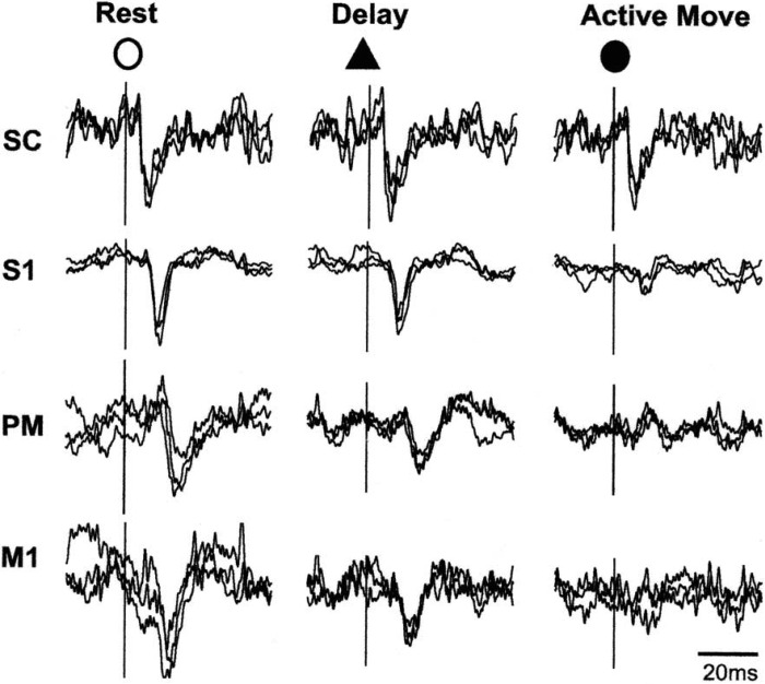
Fig. 6.
Superimposed responses recorded from cervical spinal cord (SC), primary somatosensory cortex (S1), premotor cortex (PM), and primary motor cortex (M1) following stimulation of the superficial radial nerve in a monkey. During active flexion of the wrist the cortical responses are markedly diminished from the resting values, and there is also some reduction in the motor and premotor areas when the animal is waiting for the signal to contract. Reproduced from Seki and Fetz (2012), with permission of K. Seki.

