Fig. 2.
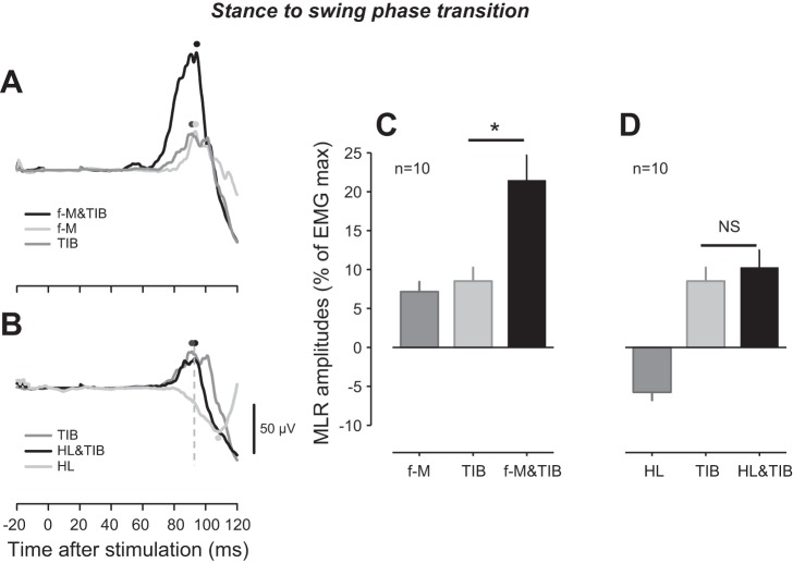
Reflex modulation of middle latency response (MLR) following tibial nerve (TIB), forefoot-medial (f-M), forefoot-lateral (f-L), heel (HL), and combined f-M&TIB and HL&TIB stimulation at the stance-to-swing-phase transition. A: superimposed TA EMG recordings following f-M, TIB, and f-M&TIB stimulation obtained from a single subject. B: EMG recordings of TA following HL, TIB, and HL&TIB stimulation, obtained from the same subject showed in A. C: group means of MLR amplitudes following f-M, TIB, and f-M&TIB stimulation. D: group means of MLR following HL, TIB, and HL&TIB stimulation. Note that the EMG traces for TIB stimulation in A and B are the same; furthermore, the group data for TIB in C and D are the same. Black dots, peak latency of MLR following f-M&TIB (A) and HL&TIB (B) stimulation; dark-gray dots, peak latency of MLR following TIB stimulation (A and B); light-gray dots, peak latency of MLR following f-M (A) and HL (B) stimulation; dashed, vertical line (B), latency of peak amplitude following HL&TIB stimulation. *P < 0.01; NS, not significant.

