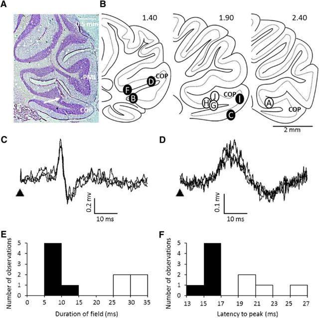
Figure 1.
Cerebellar localization and nature of hindlimb evoked fields. A, Example case showing end of electrode track (arrows) within the copula pyramidis (COP). PML, Paramedian lobule. B, Standard sagittal sections of COP (lateral position from midline shown in millimeters) displaying the approximate location of electrode recording sites. Filled circles represent sites in which short-duration responses were obtained, and open circles show site of recording of long-duration responses. All sites are identified with the exception of animal GRE, in which the recording site could not be recovered histologically. C, Example extracellular recordings (3 sweeps superimposed) showing short-duration cerebellar field potential (CFP) evoked by ipsilateral hindlimb stimulation in the awake rat. Arrowhead, Time of stimulus. D, Same as C, but example of a long-duration CFP. Histogram showing the distribution of mean duration of evoked fields (E) and the mean latency to peak during rest (F) (n = 10 rats).

