
Figure 6.
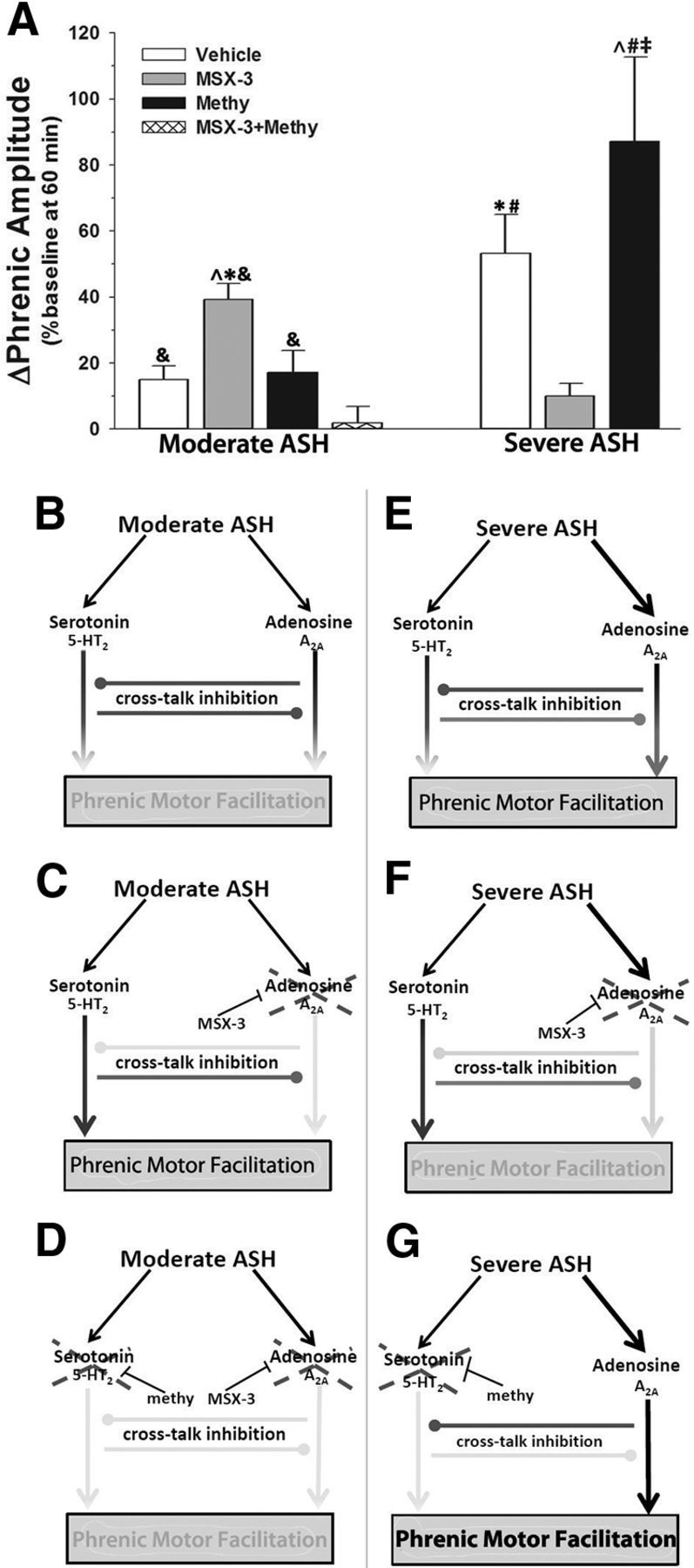
Competing 5-HT2/A2A-dependent mechanisms could explain pattern sensitivity of pMF induced by moderate hypoxia. A, Summary of pMF following mASH or sASH in rats treated with vehicle, 200 mm MSX-3, 20 mm methysergide, or 20 mm methysergide + 200 mm MSX-3. B, Proposed 5-HT2/A2A interactions predict magnitude of pMF. ↓ indicates activation. Gray line with gray circle represents inhibition. Gray line with intersecting bar represents blockade by intrathecal drug. During mASH, balanced activation of 5-HT2- and A2A-dependent pathways allows for cross talk inhibition to constrain expression of pMF. C, Spinal blockade of A2A pathway with MSX-3 during mASH prevents cross talk inhibition of the 5-HT2 pathway, revealing pMF. D, Blockade of serotonin receptors with methysergide (methy) before A2A receptor blockade and mASH exposure prevents pMF, demonstrating that cross talk inhibition from A2A-dependent pathway restrains 5-HT2-dependent pMF during mASH. E, sASH causes pMF because of greater relative A2A receptor activation, overcoming cross talk inhibition. F, Blockade of spinal A2A receptors prevents expression of pMF following severe ASH, demonstrating that pMF following severe ASH is A2A-dependent. G, Blockade of spinal serotonin receptors prevents cross talk inhibition of the A2A pathway during severe ASH, causing enhanced pMF. *Significant versus vehicle + mASH. ∧Significant versus methysergide + mASH. &Significant versus methysergide + MSX-3 + mASH. #Significant versus MX3 + sASH. ‡Significant versus vehicle + sASH.