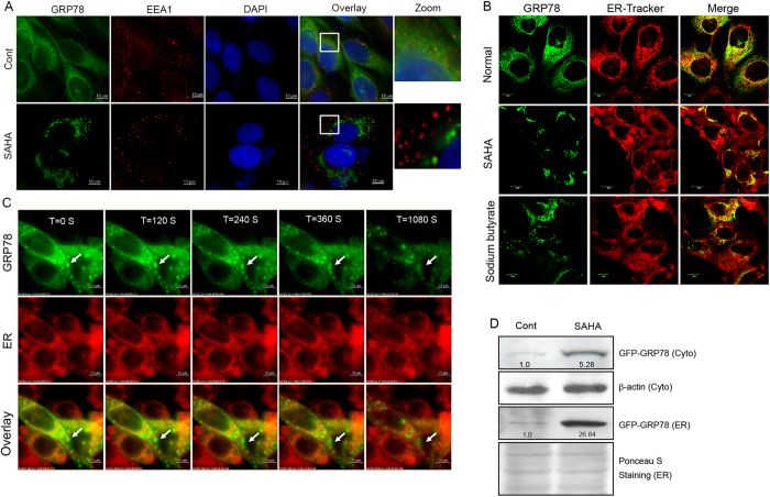
Figure 4. HDAC inhibitors induce GRP78 aggregation in the ER.
(A) DLD1 cells stably expressing GRP78-GFP were treated with SAHA and immunostained with an anti-EEA1 antibody. The nucleus was stained with DAPI. The corresponding images were superimposed to determine the degrees of colocalization. (B) DLD1 cells stably expressing GRP78-GFP were treated with SAHA or sodium butyrate. The ER was labelled with ER-Tracker Red dye, and colocalization analysis was performed using confocal microscopy. (C) Immunofluorescence photomicrographs of GRP78-GFP-expressing DLD1 cells treated with SAHA at different time points (120, 240, 360, 1080 s). (D) ER fractions were isolated from GRP78-GFP-expressing cells with or without SAHA treatment for 24 h. After cell lysis, ER and cytoplasmic fractions were subjected to Western blot analysis to probe for GRP78-GFP.

