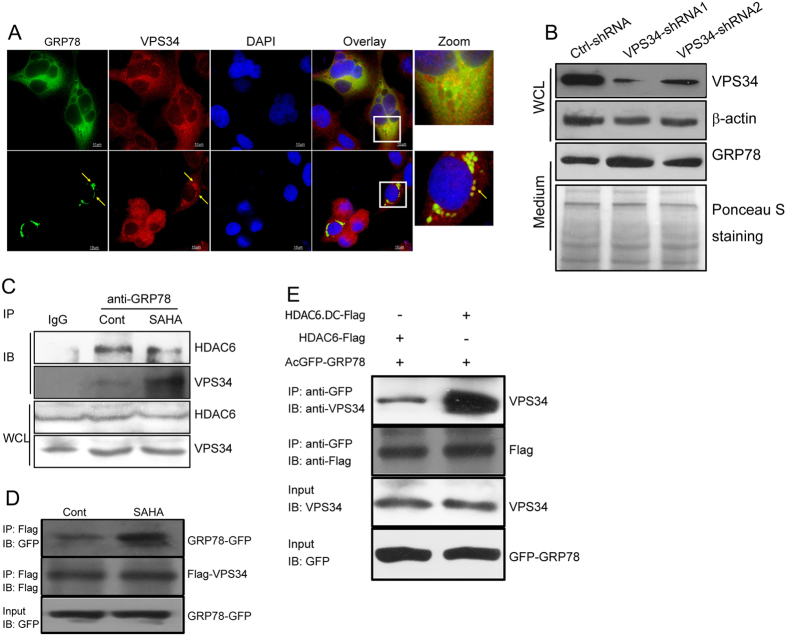
Figure 6. The aggregation of GRP78 is associated with VPS34.
(A) DLD1 cells stably expressing GRP78-GFP were treated with or without SAHA and immunostained with an anti-VPS34 antibody. The images were superimposed to determine the degree of colocalization. (B) VPS34 expression in DLD1 cells was knocked down by shRNAs. Culture supernatants were collected and subjected to Western blot analysis of GRP78 secretion. (C) DLD1 cells treated or untreated with SAHA were immunoprecipitated with an anti-GRP78 antibody followed by immunoblotting with anti-HDAC6 and anti-VPS34 antibodies. (D) 293T cells were transfected with Flag-tagged VPS34 and GRP78-GFP plasmids. After transfection for 24 h, cells were treated with SAHA for another 12 h. Lysates from SAHA-treated and untreated cells were immunoprecipitated with an anti-Flag antibody followed by immunoblotting with anti-GFP and anti-Flag antibodies. (E) 293T cells were co-transfected with GRP78-GFP and Flag-tagged wild-type or catalytically inactive HDAC6. Cell lysates were immunoprecipitated with an anti-GFP antibody followed by immunoblotting with anti-VPS34 and anti-Flag antibodies.

