
Figure 1. Molecular haplotyping of HLA: overview of the workflow.
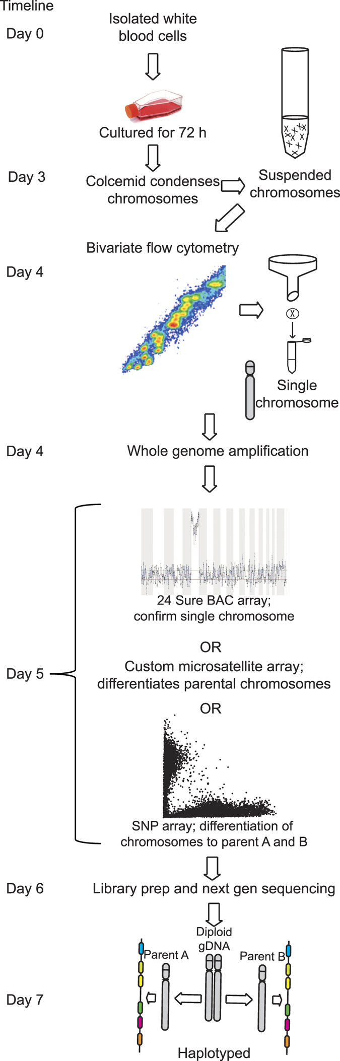
A patient’s white blood cells are purified and cultured. Addition of colcemid arrests cell division at metaphase, condensing chromosomes sufficiently to survive vigorous hypotonic cell lysis. Bivariate flow cytometry allows visual recognition of chromosome 6 which is then flow sorted into single PCR tubes. A whole genome amplification kit optimised for single cells yields sufficient material to confirm the presence of chromosome 6 via BAC and/or SNP array to discover the parental source, quality, and detection of contamination. Library generation and paired-end next-generation sequencing at 110X depth, yields sufficient coverage for HLA allele calling and haplotype phasing.