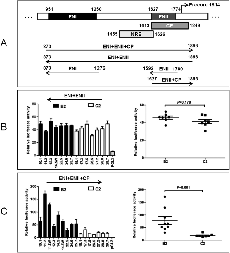
Figure 1. Schematic representation of the HBV transcriptional regulatory elements and luciferase reporter activities of a 1-kb HBV DNA fragment inserted in the sense or antisense orientation.
(A) Location of ENI, NRE, ENII, and CP in the HBV genome. The location of NRE is based on ref. 31. Also shown are the reporter constructs used in the present study. Three HBV DNA fragments were inserted to pGL3 promoter vector in the antisense orientation to measure just enhancer activities, while two DNA fragments were inserted to pGL2 basic vector in the sense orientation to determine both enhancer and promoter activities. (B) Reporter activities of a 1-kb HBV DNA fragment (873–1866) inserted in the antisense orientation to measure combined effects of ENI+NRE+ENII. (C) Reporter activities of the same DNA fragment inserted in the sense orientation to reflect combined effects of ENI+NRE+ENII+CP. The relative luciferase levels (firefly luciferase activity/renilla luciferase activity) are shown as columns in the left panels and as dots in the right panels. The P values for the difference between the B2 and C2 isolates are provided. Data shown are based on three independent transfection experiments.

