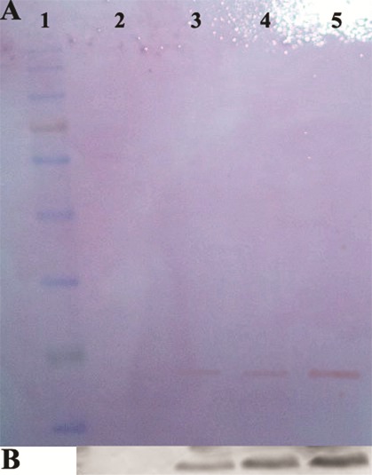
Figure 6.

Western-blot analysis with specific rabbit anti-endostatin polyclonal antibody. Panel (A) is the ponceaus staining of pvdf membrane. Lane 2 is the control sample; lane 3-5 represent the sample related to periplasmic proteins after induction, cation exchange purified rhEs and size exclusion purified rhEs, respectively. Panel (B) is the immunoblotting image of samples shown in panel A. Lanes 2, 3, 4 and 5 correspond to the lanes 2, 3, 4 and 5 in panel A. Lane 1 is protein marker.
