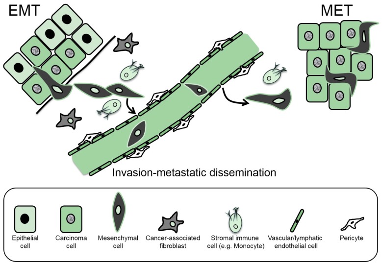
Figure 1.
Schematic representation of EMT (epithelial–mesenchymal transition) and MET (mesenchymal–epithelial transition) in the context of carcinoma progression. Hyperplastic epithelial cells (light green cytoplasm) are shown aligned along their basement membrane (thick black line). Carcinoma cells develop in this primary tumor (deep green cytoplasm) and some undergo EMT, forming mesenchymal cells (black cytoplasm) that degrade the basement membrane and invade the local microenvironment. EMT and invasiveness are enhanced by auxiliary paracrine signals from stromal fibroblasts and immune cells, thus facilitating the movement of mesenchymal cells to the blood or lymphatic vessels where they can intravasate. Upon successful survival in the lymphatic or vascular circulation, some mesenchymal cells extravasate and initiate micrometastases consisting of mesenchymal cells and a bulk of carcinoma cells (deep green cytoplasm) that are generated via MET. The cell types of the schematic are explained below the picture.

