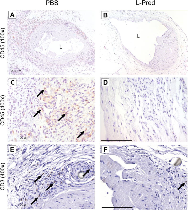
Figure 4. Representative sections of immunohistochemical stainings against CD45 and CD3 at 14 days after AVF surgery.
(A–D) CD45(+) cells (black arrow) in the venous outflow tract of the AVF. (E,F) CD3(+) cells (black arrow) in the venous outflow tract of AVF. Original magnification (A,B): 100x. Bar = 200 μm, (C–F): 400x, Bar = 100 μm (L) Lumen.

