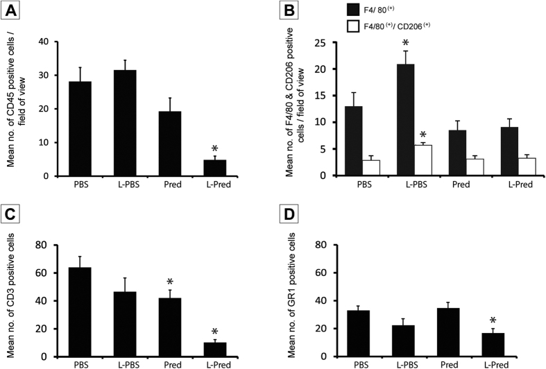
Figure 5. Quantification of immunohistological staining against different inflammatory cells at day 14.
(A) CD45(+) leucocytes. (B) F4/80(+) macrophages and F4/80(+)CD206(+) M2 macrophages. (C) CD3(+) granulocytes. (D) GR1(+) granulocytes in the venous outflow tract of AVF at day 14. (*)P < 0.05 compared to PBS.

