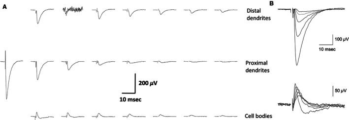
Figure 2.

Examples of simultaneous fPSPs recordings from a piriform cortex slice (A) Stimulus was applied 100 μm left to the first response on the second row (layer Ib). Note the rapid decline in fPSP amplitude as the distance from the point of stimulation increases. Also, note that while the responses in the two upper rows, which represent recordings from apical dendrites are negative going, indicating inward synaptic currents, responses in the cell body layer are positive, indicating outward currents. Recording are shown in 22 out of 32 recording electrodes, where fPSPs are detected. Slice was taken from a pseudotrained rat. (B) Responses to a set of increasing stimulus intensity recorded from the second electrode in the second row and the first electrode from the third row (the first column from the left). The amplitude of both fPSP increases with the increase in stimulus intensities, but inward synaptic currents are maintained restricted to the dendrites.
