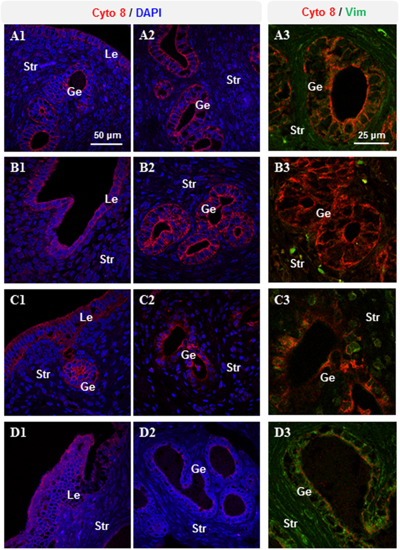
Figure 3. Immunofluorescence detection of cytokeratin 8 and vimentin in rats treated with insulin and/or hCG.
Representative images are shown. The investigators were blinded to allocation for immunofluorescence analyses (n = 10/group). Lu, lumen; Le, luminal epithelial cells; Ge, glandular epithelial cells; Str, stromal cells. Scale bars are indicated in the photomicrographs.

