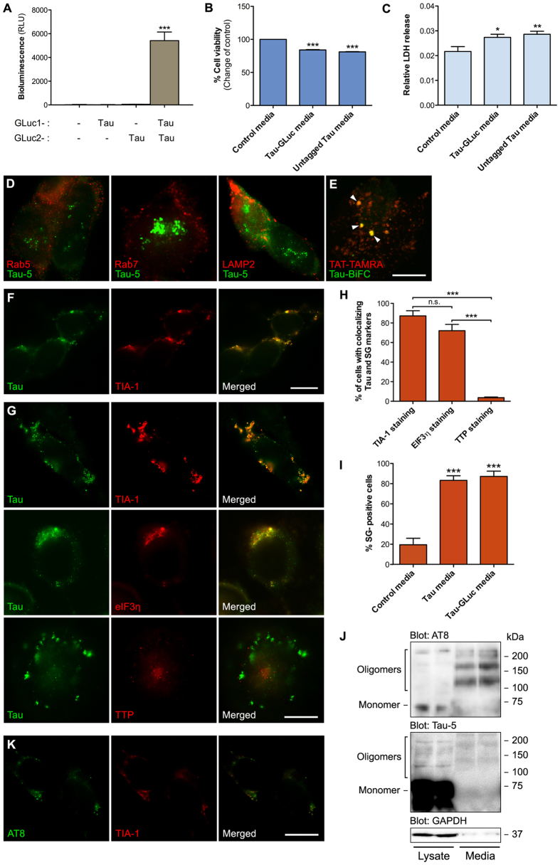
Figure 2. Internalized extracellular Tau localizes to stress granules.
(A) PCA from conditioned media collected from HEK293T transiently transfected with either mock plasmid, Tau-GLuc1, Tau-GLuc2 or Tau-GLuc1/Tau-GLuc2. Secreted Tau dimers accumulate in fresh serum-free media (n = 3). Media was conditioned for 24 h with HEK293T cells expressing different forms of Tau for the subsequent experiments. (B) Resazurin-based cell viability assay showing decreased viability of cells exposed to Tau-GLuc1/2-containing media compared to the control-media exposed cells (n = 6). (C) Relative LDH release for cells exposed to Tau-GLuc1/2-containing media show slightly decreased viability compared to the control cells (n = 4). (D) Naïve HEK293T cells were exposed to Tau-GLuc conditioned media for 6 h and stained with Tau-5 and (from left to right) Rab5, Rab7 and Lamp2 antibodies to mark early endosomes, late endosomes and lysosomes, respectively. (E) Fluorescent TAT-TAMRA peptide was used as a marker for macropinosomes and visualized in live-cell imaging together with Tau-BiFC containing media, showing some colocalization. Tau-BiFC media was produced collecting conditioned media from HEK293T cells transfected with Tau-GFP10C and Tau-GFP11C, two Tau constructs carrying complementary fragments of GFP. Similar to GLuc-based PCA, generation of GFP signal requires Tau dimerization. (F) Naïve HEK293T cells were exposed to untagged Tau conditioned media for 6 h and stained with Tau-5 and SG marker TIA-1. (G) Naïve HEK293T cells exposed to Tau-GLuc1/2 containing media for 6 h and costained with Tau-5 and SG markers TIA-1, eIF3η and TTP. (H) Quantitative analysis of SGs using different markers indicates that TIA-1 and eIF3η staining frequently localize with Tau while TTP colocalization with Tau is found in very few cells (n = 4). (I) Quantitative analysis of TIA-1-positive SG formation in cells conditioned with Tau-GLuc and Tau media compared to control media (n = 4). (J) Western blot of Tau-GLuc-transfected cell lysate and Tau-GLuc conditioned media, stained with AT8, Tau-5 and GAPDH antibodies. Media was concentrated by 250x using filter centrifugation before loading on gel. The blot picture was cropped from a larger image, maintaining all the stained bands. (K) Naïve HEK293T cells were exposed to Tau-GLuc conditioned media for 6 h and stained with AT8 and TIA-1 antibodies, showing colocalization of Ser199/Ser202/Thr205-phosphorylated Tau and TIA-1 in the recipient cells. Scalebar = 10 μm; average +/−SEM is shown; ***p < 0.001; **p < 0.005; *p < 0.05.

