
Figure 4. Internalized Tau is recruited to stress granules in N2A cells.
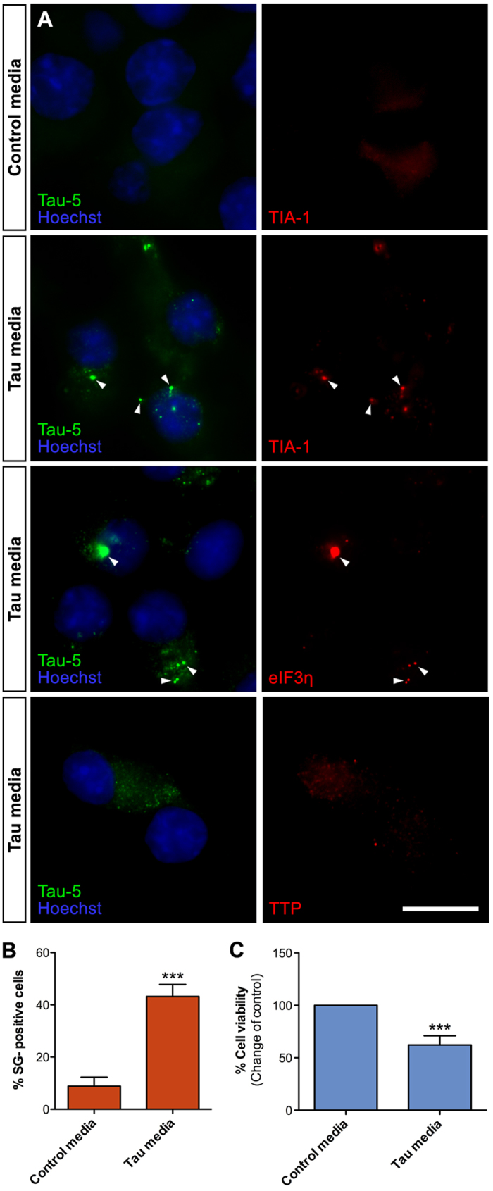
(A) Naïve N2A cells exposed to Tau-GLuc conditioned media for 6 h and stained with Tau-5 and stress granule markers TIA-1, eIF3η and TTP. TIA-1 and eIF3η show clear colocalization with internalized Tau, while TTP does not show any. (B) Quantitative analysis of SG formation in cells exposed to Tau conditioned media compared to control media indicates that about 40% of N2A cells contained TIA-1-positive SGs following Tau uptake (n = 4). (C) Resazurin-based cell viability assay indicates that viability of N2A cells exposed to Tau-GLuc media was significantly decreased compared to cells exposed to control media (n = 8). Scalebar = 10 μm; average +/−SEM is shown; ***p < 0.001.