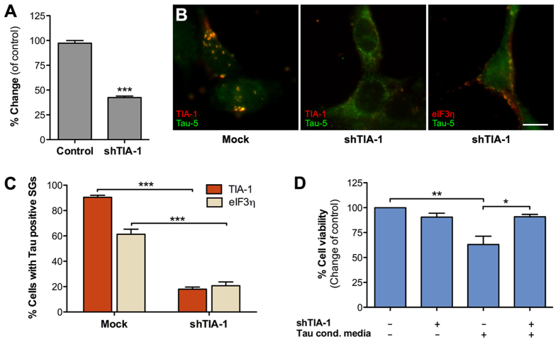
Figure 5. Tau localization to stress granules requires TIA-1.
(A) TIA-1 shRNA knockdown efficiency determined by qPCR following shRNA plasmid transfection of HEK293T cells. Levels of TIA-1 mRNA were normalized to GAPDH mRNA levels (n = 2). (B) TIA-1 was transiently knocked down in HEK293T cells (middle and right images) that were then exposed to Tau-GLuc-conditioned media and stained with Tau-5 and stress granule marker antibodies. The middle image shows typical cells with mostly cytosolic, non-punctate staining of Tau. The image on the right shows an example of a cell with SGs costaining with Tau and eIF3η. (C) Quantitative analysis of Tau-positive stress granule formation. SGs were present in the majority of cells exposed to Tau media when TIA-1 is normally expressed but were significantly decreased when TIA-1 was knocked down. Similar results were obtained with both TIA-1 and eIF3η staining (n = 3). (D) Resazurin-based cell viability assay showed that TIA-1 knockdown had no effect on cell viability per se but was able to improve viability in cells exposed to Tau-GLuc-conditioned media (n = 3). Scalebar = 10 μm; average +/− SEM is shown; ***p < 0.001; **p < 0.01; *p < 0.05.

