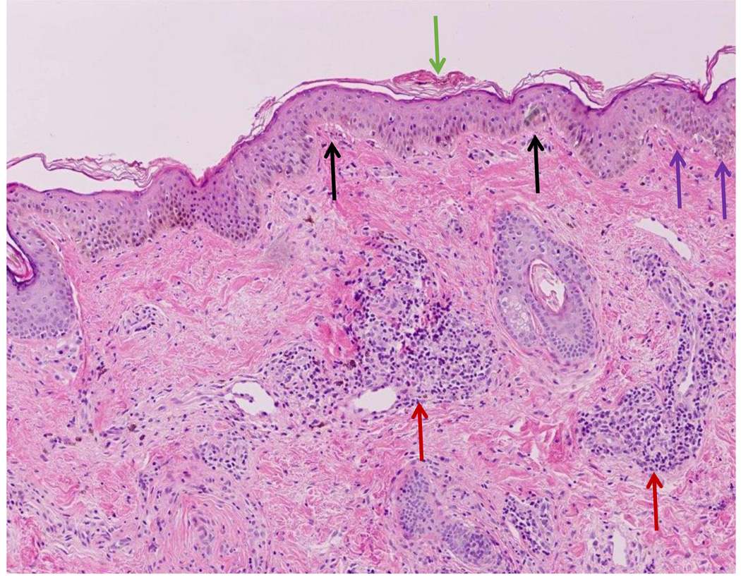
Figure 1.
Classic histopathology for discoid lupus erythematosus is demonstrated including hyperkeratosis of the stratum corneum (green arrow), mild thickening of the basement membrane (purple arrows), and vacuolar interface dermatitis at the dermal-epidermal junction (black arrows). There are also lymphoid infiltrates surrounding both blood vessels and eccrine glands in the dermis (red arrows).

